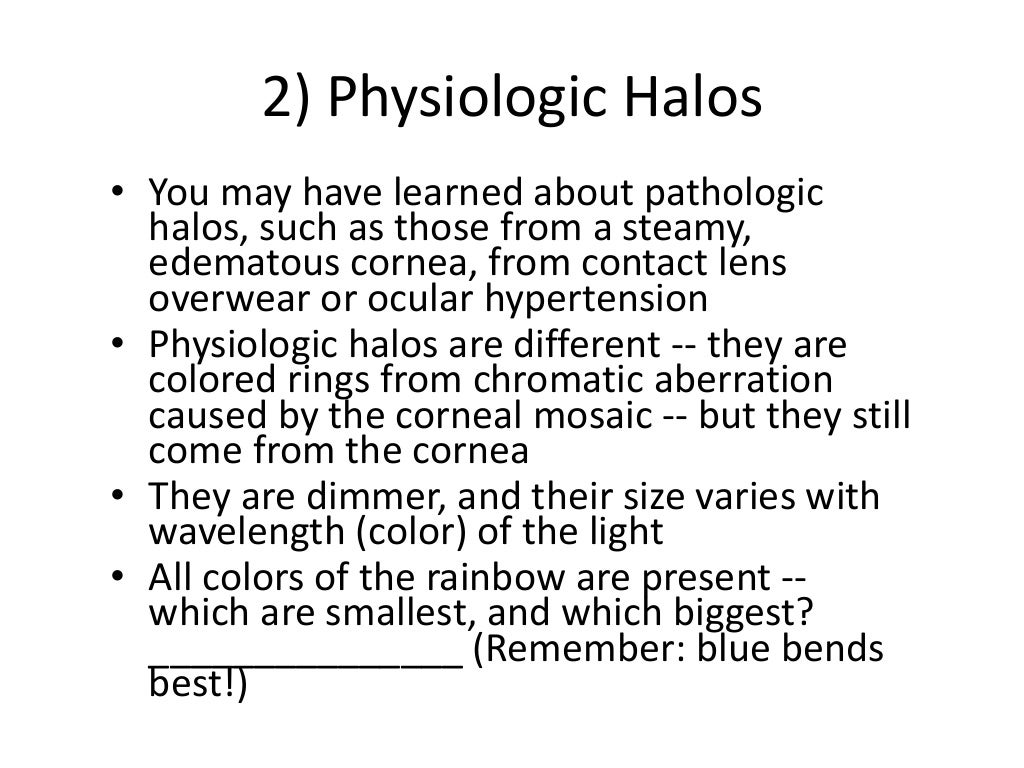
A smaller halo (diameter of approximately 3 degrees) is a result of the structure and organization of the corneal epithelial cells, whereas the corneal endothelial cells and/or lens epithelial cells contribute to a slightly larger halo (approximately 4.5 degrees in diameter). 4 A larger halo (approximately 9 degrees) was first described by Descartes in 1637 and appears to be caused by second-order diffraction by the corneal endothelium. This produces a halo with diffraction rings that have been estimated to have diameters ranging from 4.5 degrees for violet, 5 degrees for blue, 5.4 degrees for green, and 6 degrees for yellow. In this case, the halo that generally is most visible results from diffraction by the radially arranged lens fibers. With a dilated pupil, the effect of the peripheral lens becomes marked. Because the axial portion of the lens appears to be relatively uniform, no lenticular halo is observed with a pupil less than 3 mm in diameter. As a result, the crystalline lens may be regarded as a strong positive lens with a radial diffraction grating (similar to any two- or three-dimensional array of lines or dots showing periodic variations of either transparency or refractive index) superimposed on the periphery. 2, 3 With the exception of the zonular insertions and anterior cellular layer, the crystalline lens is composed of fibers that pass in an approximately radial manner from the anterior to the posterior sutures. In the healthy eye there are differences in size between halos associated with different ocular structures. Halos can result from either the normal physical properties of ocular structures or the alterations in these physical properties caused by the response of ocular tissues to pathology. Halos are smaller when the diffracting structures are nearer to the retina. The perceived diameter of an entoptic halo also depends on the distance between the diffracting structures and the retina. The longer the wavelength of light, the larger the diameter of the halo. Therefore, as the diameter of the diffracting structures increases (e.g., cells, droplets) or the spacing of the elements in the diffraction grating gets larger (e.g., lens fibers) the diameter of the halo will become smaller. Diffraction theory predicts that the angular diameter of a halo is related directly to the wavelength of the light and inversely to the size (or in the case of a regular pattern to the spacing) of the diffracting structure. The specific characteristics of individual halos depend on the structure of the diffraction grating underlying their generation. Halos are caused by diffraction from small regularly sized or spaced structures within the eye. With a white light source, the outside rings of the diffraction spectra are red, while the inner rings are blue–violet. Entoptically, these diffraction spectra are visible as rainbow-like halos or coronas centered on the source. Ocular structures that act as diffraction gratings produce diffraction spectra when a point source is viewed. The closer the opacity lies to the pinhole, the greater the relative displacement. This is perceived as if the shadow moved in the same direction as the image of the pupil. As a result, the shadow of the opacity is no longer centered in the entoptic image of the pupil. Once again when the pinhole is shifted downward, both the entoptic image of the pupil ( green lines) and the shadow of the opacity ( yellow shadow) are displaced upward, but in this case there is relatively more displacement of the shadow. In the second case (B), the opacity is positioned anterior to the entrance pupil of the eye. The closer the opacity lies to the retina, the greater the relative displacement.


This is perceived as if the shadow moved in the opposite direction from the image of the pupil. When the pinhole is shifted downward, both the entoptic image of the pupil ( green lines) and the shadow of the opacity ( yellow shadow) are displaced upward, but there is relatively less displacement of the shadow.

In the first case (A), the opacity is positioned posterior to the entrance pupil of the eye. In both cases (A and B), the shadow of the opacity ( black shadow) appears centered within the entoptic image of the pupil ( red lines) when the pinhole is positioned on the optic axis. In the two cases illustrated here, a pinhole source is shifted downward from the optical axis (as indicated by the arrow), and the amount and direction of the displacement of the shadow of an opacity relative to the entoptic image of the pupil is evaluated. 1 The use of relative entoptic parallax to estimate the position of an opacity in the eye.


 0 kommentar(er)
